20191219h Day -12: Influenza Immunity and Vaccines

image
3D computer-generated rendering of an influenza virus. (Credit: Dan Higgins, courtesy of CDC/ Douglas Jordan)

20200520W Santa Cruz, CA: The first article to catch my eye today as I looked back to Dec 19, 2019, is Why your first battle with flu matters most, a article from the University of Arizona about a research paper published online on this same date: Childhood immune imprinting to influenza A shapes birth year-specific risk during seasonal H1N1 and H3N2 epidemics.

The researchers looked at Influenza virus subtypes H1N1 and H3N2 and found that “birth year-specific differences in childhood immune imprinting, not differences in evolutionary rate, explain differences in H1N1 and H3N2’s age-specific impacts.”

A result found by the researches was evidence to support the view that immune protection is stronger when acquired as a child rather than as an adult:

The fact that elderly cohorts show relatively weak immune protection against H3N2, even after living through decades of seasonal exposure to or vaccination against H3N2, suggests that antibody responses acquired in adulthood do not provide the same strength or durability of immune protection as responses primed in childhood.

Another possible conclusion not mentioned is that the H3N2 vaccinations, versus naturally acquired immunity, may partially explain the reduced immune protection in adults.

The University of Arizona article has some interesting quotes:

“Clearly, something compromises the immunity to strains that you see secondarily, even if they belong to the same group as your first exposure,” Worobey adds. “The second subtype you’re exposed to is not able to create an immune response that is as protective and durable as the first.”

In other words, our ability to fight off the flu virus is determined not only by the subtypes we have encountered over the course of our lives, but also by the sequence in which we have encountered them.

“Whichever subtype our immune system sees first lays down an imprint that protects us especially well against strains of the same subtype,” Worobey says, “but relatively poorly against strains from other subtypes, even though you’ve encountered those subsequently.”

The molecular causes of this effect are currently being studied, according to the researchers.

“Part of your immune system’s response to current infection is directed against the strain you first had as a kid, and that investment of fighting the last war appears to compromise your ability to form a fully effective immune response to the invader you encounter later,” Worobey says.

Regarding vaccines, Worobey, one of the researchers, has this to say:

“We need a vaccine that targets the deficits on an individualized level,” Worobey says. “Our work has clearly shown that the first virus we had can have a profound long-term effect. The bad side of that is that our immune system seems to be locked into fighting just one half of flu genetic diversity, and we need to find ways of breaking that.”

This research suggest to me that if we keep SARS-CoV-2 from infecting our children today, then they will be more susceptible to infection from SARS-CoV-2 even decades from now. It also makes sense to me that vaccines should not be the first solution thought of when deciding how to protect the population from a pathogen, and perhaps should be the last solution considered. Instead, the development of anti-virals that can assist the immune system in fighting off a viral pathogen make more sense. This would allow the body to naturally develop an immunity. Also, we need to develop treatments to improve recovery time and protect the body from the long-term effects of a particular viral infection.

20191220F Day -11: Human Coronaviruses and the Central Nervous System

20200520W Santa Cruz, CA: When I searched back 5 months to see what was going on related to the COVID-19 pandemic, I found a research paper published online on Dec. 20, 2019: Human Coronaviruses and Other Respiratory Viruses: Underestimated Opportunistic Pathogens of the Central Nervous System?

Since the effect of COVID-19 on the CNS has also been discussed lately, I thought it would be interesting to go through this paper and see what quotes leap out at me. So here goes.

First from the Abstract, some key points so you know what you’re getting into. This is the entire Abstract from the paper in bullet form so that I can easily refer back to it later, if needed.

  1. Respiratory viruses infect the human upper respiratory tract, mostly causing mild diseases. However, in vulnerable populations, such as newborns, infants, the elderly and immune-compromised individuals, these opportunistic pathogens can also affect the lower respiratory tract, causing a more severe disease (e.g., pneumonia).
  2. Respiratory viruses can also exacerbate asthma and lead to various types of respiratory distress syndromes.
  3. Furthermore, as they can adapt fast and cross the species barrier, some of these pathogens, like influenza A and SARS-CoV, have occasionally caused epidemics or pandemics, and were associated with more serious clinical diseases and even mortality.
  4. For a few decades now, data reported in the scientific literature has also demonstrated that several respiratory viruses have neuroinvasive capacities, since they can spread from the respiratory tract to the central nervous system (CNS). Viruses infecting human CNS cells could then cause different types of encephalopathy, including encephalitis, and long-term neurological diseases.
  5. Like other well-recognized neuroinvasive human viruses, respiratory viruses may damage the CNS as a result of misdirected host immune responses that could be associated with autoimmunity in susceptible individuals (virus-induced neuro-immunopathology) and/or viral replication, which directly causes damage to CNS cells (virus-induced neuropathology).
  6. The etiological agent of several neurological disorders remains unidentified. Opportunistic human respiratory pathogens could be associated with the triggering or the exacerbation of these disorders whose etiology remains poorly understood.
  7. Herein, we present a global portrait of some of the most prevalent or emerging human respiratory viruses that have been associated with possible pathogenic processes in CNS infection, with a special emphasis on human coronaviruses.

Here are some interesting facts quoted from the Introduction:

  1. Considering all types of viral infections, between 6000 and 20,000 cases of encephalitis that require hospitalization occur every year in the United States, representing about 6 cases per 100,000 infected persons every year.
  2. Viruses represent the most prevalent pathogens present in the respiratory tract. Indeed, it is estimated that about 200 different viruses (including influenza viruses, coronaviruses, rhinoviruses, adenoviruses, metapneumoviruses, such as human metapneumovirus A1, as well as orthopneumoviruses, such as the human respiratory syncytial virus) can infect the human airway.
  3. new respiratory viral agents emerge from time to time, causing viral epidemics or pandemics associated with more serious symptoms, such as neurologic disorders. These peculiar events usually take place when RNA viruses like influenza A, human coronaviruses, such as MERS-CoV and SARS-CoV, or henipaviruses, present in an animal reservoir, cross the species barrier as an opportunistic strategy to adapt to new environments and/or new hosts.

In the rest of the paper, I found these quotes of interest:

Respiratory viruses such as RSV, henipaviruses, influenza A and B, and enterovirus D68 are also sometimes found in the blood and, being neuroinvasive, they may therefore use the hematogenous route to reach the CNS.


Influenza viruses are classified in four types: A, B, C and D. All are endemic viruses with types A and B being the most prevalent and causing the flu syndrome, characterized by chills, fever, headache, sore throat and muscle pain. They are responsible for seasonal epidemics that affect 3 to 5 million humans, among which 500,000 to 1 million cases are lethal each year. Associated with all major pandemics since the beginning of the 20th century, circulating influenza A presents the greatest threat to human health.


Last but not least, human coronaviruses (HCoV) are another group of respiratory viruses that can naturally reach the CNS in humans and could potentially be associated with neurological symptoms. These ubiquitous human pathogens are molecularly related in structure and mode of replication with neuroinvasive animal coronaviruses.

Taken together, all these data bring us to consider a plausible involvement of HCoV in neurological diseases.

As I read the following, I’m struct by how it could be written about the current SARS-2 (COVID-19) pandemic. Even the CFR of 10% is not that far off if there is only limited testing. I’m curious now to know if serological testing of SARS-1 has been done in SARS-1 outbreak areas to determine the true infection spread in those areas.

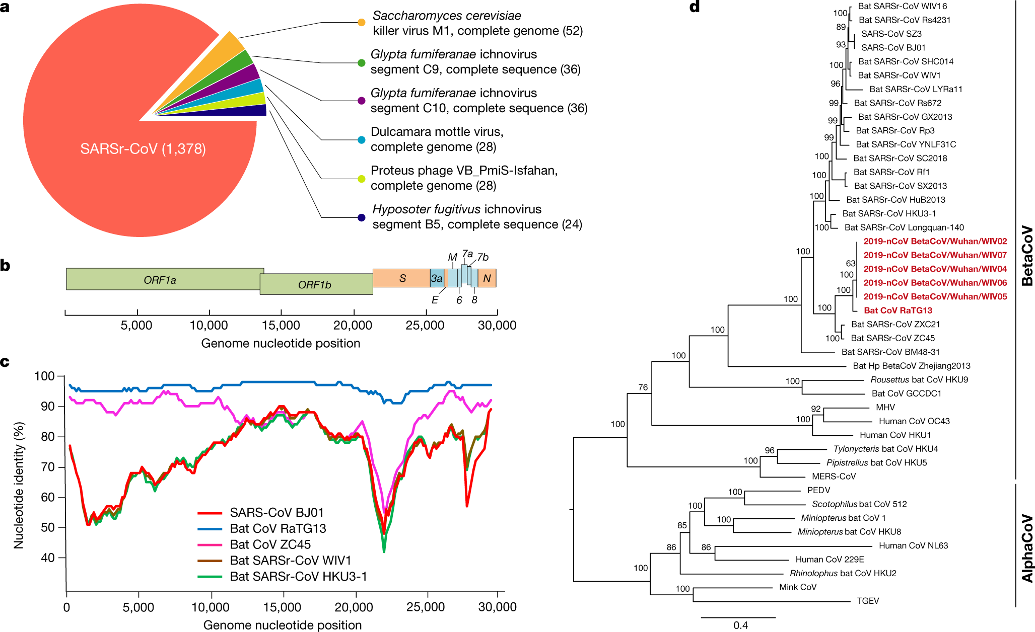
The 2002–2003 SARS pandemic was caused by a coronavirus that emerged from bats (first reservoir) to infect palm civets (intermediary reservoir) and then humans . A total of 8096 probable cases were reported and almost 10% (774 cases in more than 30 countries) of these resulted in death. The clinical portrait was described as an initial flu-like syndrome, followed by a respiratory syndrome associated with cough and dyspnea, complicated with the “real” severe acute respiratory syndrome (SARS) in about 20% of the patients. In addition, multiple organ failure was observed in several SARS-CoV-infected patients .

And the following “inefficient human-to-human transmission” and “more efficient human-to-human transmission in S. Korea” mentioned below shows how both of these universes can exist.

Although possible, human-to-human MERS-CoV transmission appears inefficient as it requires extended close contact with an infected individual. Consequently, most transmission have occurred among patients’ families and healthcare workers (clusters of transmission). A more efficient human-to-human transmission was observed in South Korea, during the 2015 outbreak of MERS-CoV. Even though it has propagated to a few thousand people and possesses a high degree of virulence, MERS-CoV seems mostly restricted to the Arabic peninsula and is not currently considered an important pandemic threat. However, virus surveillance and better characterization are warranted, in order to be prompt to respond to any change in that matter.

As I read the following about SARS-1 and MERS, the same populations seem to be infected except that not much is reported about SARS-2/COVID-19 affecting infants.

As of October 8, 2019, the World Health Organization (WHO) reported that MERS-CoV had spread to at least 27 different countries, where 2468 laboratory-confirmed human cases have been identified with 851 being fatal (https://www.who.int/emergencies/mers-cov/en/). As observed for the four circulating strains of HCoV, both SARS-CoV and MERS-CoV usually induce more severe illnesses, and strike stronger in vulnerable populations such as the elderly, infants, immune-compromised individuals or patients with comorbidities.

A comparison between the endemic coronavirus, HCoV-OC43, and both SARS-CoV-1 and SARS-CoV-2 seems like it would be helpful in better understanding these coronaviruses.

After an intranasal infection, both HCoV-OC43 and SARS-CoV were shown to infect the respiratory tract in mice and to be neuroinvasive. Over the years, we and others have gathered data showing that HCoV-OC43 is naturally neuroinvasive in both mice and humans.

Here’s a mention of the viral glycoprotein (S):

Immune cell infiltration and cytokine production were observed in the mouse CNS after infection by HCoV-OC43. This immune response was significantly increased after infection by viral variants, which harbor mutations in the viral glycoprotein (S).

Virus–cell interactions are always important in the regulation of cell response to infection. For HCoV-OC43, we clearly showed that the viral S and E proteins are important factors of neurovirulence, neuropropagation and neurodegeneration of infected cells.

And this on Hemagglutinin-esterase (HE protein) seems of interest:

We have also demonstrated that the HE protein is important for the production of infectious HCoV-OC43 and for efficient spreading between neuronal cells, suggesting an attenuation of the eventual spread into the CNS of viruses made deficient in fully active HE protein, potentially associated with a reduced neurovirulence.

This final paragraph sums up the risk of chronic human neurological diseases tied to coronavirus infections.

Like for several other respiratory viruses, accumulating evidence now indicate that HCoV are neuroinvasive in humans and we hypothesize that these recognized respiratory pathogens are potentially neurovirulent as well, as they could participate in short- and long-term neurological disorders either as a result of inadequate host immune responses and/or viral propagation in the CNS, which directly induces damage to resident cells. With that in mind, one can envisage that, under the right circumstances, HCoV may successfully reach and colonize the CNS, an issue largely deserted and possibly underestimated by the scientific community that has impacted or will impact the life of several unknowing individuals. In acute encephalitis, viral replication occurs in the brain tissue itself, possibly causing destructive lesions of the nervous tissue with different outcomes depending on the infected regions. As previously mentioned, HCoV may persist in the human CNS as it does in mice and potentially be associated with different types of long-term sequelae and chronic human neurological diseases.

The conclusions specifically calls out human coronaviruses. It also states the belief that Koch’s postulates should be modified to account for cases where diseases result rarely from a prior infection. It also refers to Multiple Schlerosis.

Several human respiratory viruses are neuroinvasive and neurotropic, with potential neuropathological consequences in vulnerable populations. Understanding the underpinning mechanisms of neuroinvasion and interaction of respiratory viruses (including HCoV) with the nervous system is essential to evaluate potentially pathological short- and long-term consequences. However, viral infections related to diseases that are rare manifestations of an infection (like long term chronic neurological diseases), represent situations where Koch’s postulates [] need to be modified. A series of new criteria, adapted from Sir Austin Bradford Hill, for causation [,] was elaborated by Giovannoni and collaborators concerning the plausible viral hypothesis in MS [].

The feeling I’m having now is to understand the fear that anti-vaccine folks have when presented with the idea of giving everyone in the world a “limited” viral infection in order to provide everyone with protective antibodies.


20191224T Day -7: Dried Cacao and SARS-CoV-2 share visual similarity

20191223-Cocao-Mystery-Shapes-Bottom-Mug
Mysterious shapes at the bottom of my cacao mug – Austin, Texas – Dec 23, 2019

20200514h Santa Cruz, CA: On the 24th of December, 2019, a bronchoalveolar lavage fluid sample from a patient with influenza of unknown origin was sent from Wuhan Central Hospital to Vision Medicals in Guangzhou. Vision Medicals is a private company that specializes in meta genomic massive parallel sequencing analysis. Three days later, Vision Medicals notified Wuhan Central Hospital that the sample contained a novel coronavirus (Reference: Wikipedia Pandemic Timeline).

What does this have to do with the mysterious shapes at the bottom of my cacao mug? Well, in looking more closely at the full shape, I now see a spherical shape that resembles a coronavirus. Compare with this image of SARS-CoV-2:

image
SARS-CoV-2

I found the SARS-CoV-2 image in a May 4th, 2020 article from Cornell by Krishna Ramanujan: Structure of COVID-19 virus hints at key to high infection rate. The news article reviews some findings from a May 1st, 2020 paper in the Journal of Molecular Biology: Phylogenetic Analysis and Structural Modeling of SARS-CoV-2 Spike Protein Reveals an Evolutionary Distinct and Proteolytically Sensitive Activation Loop.

An interesting quote from this paper is:

Structural modeling of SARS-CoV-2 S reveals a proteolytically sensitive loop

The alignment in Figure 3(b) shows a four-amino-acid insertion 681PRRA684, as well as a conserved arginine corresponding to R685 at the S1/S2 site of the SARS-CoV-2. This insertion, which appears to be unique among lineage B betacoronaviruses, suggests a differential mechanism of activation for the SARS-CoV-2 compared to other SARS-CoV and SARS-like BatCoV.

and in the Discussion section:

In this study, we show the presence of a distinct insert that maps to the S1/S2 priming loop of the SARS-CoV-2 spike protein and is not shared with SARS-CoV or any SARS-related viruses in Betacoronavirus lineage B.

20200512T Day 133: Year of the RaTG13 – Origin of SARS-CoV-2 In the Multiverse

Screen Shot 2020-05-12 at 7.58.11 PM
From Ang Ku Kueh Girl and Friend’s Chinese New Year sticker pack

20200512T Santa Cruz, CA: In this post, I’ll be discussing the origins of the coronavirus known as SARS-CoV-2 in the different sets of universes around us. Some of these universes will match the official view and some of these universes will align with conspiracy theories. Most of these universes will interfere with one another and have overlapping characteristics.

The first statement out of China was that SARS-CoV-2 was believed to have originated at a wet market in Wuhan, a large city in Hubei Province, China. Many of the early cases of COVID-19, the disease caused by SARS-CoV-2, were found in individuals who shopped at Huanan Seafood Wholesale Market. The market was closed on Jan 1, 2020, and any evidence of origin was believed to have been destroyed. A few days ago, a WHO scientist Dr. Peter Ben Embarek stated that samples taken from the wet market show that the market likely played a role in the COVID-19 outbreak. Specifically, he stated:

The market played a role in the event, that’s clear. But what role we don’t know. Whether it was the source or amplifying setting or just a coincidence that some cases were detected in and around that market,” said Dr Peter Ben Embarek in a press briefing.

Notice how this statement does not limit us to universes in which SARS-CoV-2 originated in the wet market, but also includes universes where it amplified the spread of COVID-19 and also universes in which it played no role and was only coincidentally connected.

The main unofficial and so-called conspiracy theory is that SARS-CoV-2 originated in the Wuhan Biolab not far from the wet market. This theory is circumstantial based mainly on the fact that the biolab does perform research on bat coronaviruses. This theory has two sub-theories – one in which the virus is a bioweapon and one in which it is a natural virus. There are also two overlapping sub-theories – one in which the virus was intentionally released and one in which it was accidentally released. The accidentally released theory can be further divided into one in which a lab worker is accidentally infected and one in which samples are improperly discarded.

If we assume that there is a set of universes for each of these theories and sub-theories, then the reality we see will likely be an interference pattern of these different sets of universes until one set reveals itself.

Let’s make the hypothesis/assumption that sets of universes that are in conflict will repel each other and sets of universes that are congruent will attract one another. We can begin to see a set of universes that fit together with the following features:

  1. The Huanan Seafood Wholesale Market, a wet market, played a role in the outbreak.
  2. The Wuhan Institute of Virology (WIV), a Biosafety Level 4 Laboratory, played a role in the outbreak.
  3. SARS-CoV-2 was a coronavirus being studied at WIV.
  4. SARS-CoV-2 found a way from WIV to individuals at the wet market, which was the first identified outbreak.

There are also universes where the WIV is not involved and SARS-CoV-2 is found to have come from livestock sold at the wet market. However, the future in which this is true does not appear to be sending any indications to the present that are detectible. It doesn’t fit in well with the other universes. If SARS-CoV-2 is found in live animals, it is most likely in bats not local to the Wuhan area and most likely bats from which virus samples had been collected by lab workers at WIV.

Since the SARS-CoV-2 coronavirus has been sequenced, two other coronavirus sequences have been released. The first one released is labeled RaTG13, from a sample collected in 2013 in the Yunnan province. Comparison between RaTG13 (MN996532.1) and SARSCoV2-Wuhan-Hu1 (MN908947.3) show that they had a most recent common ancestor estimated at 50 years ago. The second one released is labeled RmYN02, which is shown to have a most recent common ancestor estimated at 35 years ago. Neither one of these is likely to be the natural ancestor of the virus, due to the number of mutations between each and SARS-CoV-2.

The Bat coronavirus RaTG13 complete genome sequence references the Nature paper: A pneumonia outbreak associated with a new coronavirus of probable bat origin.

image
Figure 1 showing RaTG13 phylogeny (https://doi.org/10.1038/s41586-020-2012-7)

Key quotes from the paper:

Full-length genome sequences were obtained from five patients at an early stage of the outbreak. The sequences are almost identical and share 79.6% sequence identity to SARS-CoV. Furthermore, we show that 2019-nCoV is 96% identical at the whole-genome level to a bat coronavirus.

This second one I wish was explained more in how the “then found” occurred.

We then found that a short region of RNA-dependent RNA polymerase (RdRp) from a bat coronavirus (BatCoV RaTG13)—which was previously detected in Rhinolophus affinis from Yunnan province—showed high sequence identity to 2019-nCoV.

More thoughts to come…


20191229u Day -2: Amotosalen and ultraviolet A light efficiently inactivate MERS‐coronavirus in human platelet concentrates

20200509u Santa Cruz, CA: As I searched for what happened today, I came across a church shooting in Texas. For some reason, I took a picture on my phone of the video:

image_e1e9515e-2335-4262-acf5-df327e540607.img_0955
Texas Church Shooting – 2019-12-29u

The thought I had when remembering this church shooting was how the COVID-19 pandemic has affected religious services across the world.

I then searched for something else happening on this date and the first thing to catch my eye was this research paper published in the 2019-12-29 edition of Transfus Med: Amotosalen and ultraviolet A light efficiently inactivate MERS-coronavirus in human platelet concentrates. I see now that it was actually published online 2019-11-06, 11 days before the first known COVID-19 case.

Most of the paper authors are from King Abdulaziz University in Jeddah Saudi Arabia. Key quotes from the paper summary:

OBJECTIVE: This study aimed to assess the efficacy of the INTERCEPT™ Blood System [amotosalen/ultraviolet A (UVA) light] to reduce the risk of Middle East respiratory syndrome‐Coronavirus (MERS‐CoV) transmission by human platelet concentrates.

RESULTS: Complete inactivation of MERS‐CoV in spiked platelet units was achieved by treatment with Amotosalen/UVA light with a mean log reduction of 4·48 ± 0·3. Passaging of the inactivated samples in Vero E6 showed no viral replication even after nine days of incubation and three passages. Viral genomic RNA titration in inactivated samples showed titres comparable to those in pre‐treatment samples.

CONCLUSION: Amotosalen and UVA light treatment of MERS‐CoV‐spiked platelet concentrates efficiently and completely inactivated MERS‐CoV infectivity (>4 logs), suggesting that such treatment could minimise the risk of transfusion‐related MERS‐CoV transmission.

Keywords: amotosalen, MERS‐CoV, pathogen inactivation, platelets, UV

This paper is interesting because it looks at using UVA light as an antiviral, as opposed to the shorter wavelength UVC light.

Before going into the paper, I found it helpful to review UV light and where it fits into the electromagnetic spectrum. From the FDA: Ultraviolet (UV) Radiation.

image

Ultraviolet (UV) light has a frequency just a bit higher than visible light. If you remember the colors of the rainbow in order of low to high frequency using the ROYGBIV acronym, then you know the visible colors Red, Orange, Yellow, Green, Blue, Indigo, Violet. The next colors are Ultraviolet A, Ultraviolet B, and Ultraviolet C.

The longer the UV light wavelength, the more penetration light has into the Earth’s atmosphere, our skin, and our eyes. Light with wavelengths in the range 300-400 nm are blocked by the lens of our eyes. For this reason, we use 400 nm as the cut off point for defining UV light. Light with wavelengths shorter than 300 nm are blocked by the cornea or our eyes.

Ultraviolet A (UVA) is light with an ISO-recommended wavelength of 400-315 nm. Other names for UVA are “Black Light”, “Long-Wave”, or “Soft UV”. UVA light is not absorbed by the Earth’s ozone layer and passes through the atmosphere, the outer layer of our skin (epidermis), and into the middle layer of our skin (dermis).

Ultraviolet B (UVB) is light with an ISO-recommended wavelength of 315-280 nm. This medium-wave UV light is mostly absorbed by the Earth’s ozone layer. The UVB light that does pass through is absorbed by our epidermis and does not make it to our dermis. UVB light is necessary to help the epidermis produce vitamin D3.

Ultraviolet C (UVC) is light with an ISO-recommended wavelength of 280-200 nm. This short-wave UV light, also called “Hard UV”, is completely absorbed by the Earth’s ozone layer and atmosphere. Some UVC lamps operate at a wavelength of 253.7 nm, as this wavelength as been shown effective at killing or inactivating microorganisms.  “Far-UVC” light, at a wavelength of 222-207 nm, is being researched as a possible light range that will efficiently kill pathogens without harm to humans. (REF: UVC research at columbia.edu, Far-UVC light: A new tool to control the spread of airborne-mediated microbial diseases)

Vacuum UV (in another universe, it’s called UVD) is light with an ISO-recommended wavelength of 200nm-100nm.

image
REF: John L. Bezzant, M.D. (library.med.utah.edu)

Going back to the paper, it focuses on a need to be able to efficiently inactivate pathogens in the blood of blood banks as an alternative to testing. It specifically mentions the problem of blood carrying the MERS virus:

Underreporting of asymptomatic cases is presumed, and thus, the real number of infected patients is likely to be much higher than the number of reported and confirmed cases (Lessler et al., 2016), which together with mild cases may facilitate the spread of the virus and pose a possible risk for blood safety. Higher seropositivity in camel workers compared to the general population has been reported (Müller et al., 2015; Alshukairi et al., 2018). Therefore, asymptomatic donors may donate blood while they potentially carry the virus.


The authors go on to say:

The INTERCEPT™ Blood System technology inactivates a broad spectrum of pathogens, including bacteria, viruses and protozoa, in platelet concentrates prepared for transfusion (Schlenke, 2014). Using amotosalen (a photoactive compound) and UVA light, pathogens’ genomes are modified in a targeted and specific manner by cross‐linking the genomic strands, preventing transcription and replication without affecting the platelet efficacy and patient safety as demonstrated in clinical evaluations (Cid et al., 2012), national routine observations (Jutzi et al., 2018) and by haemovigilance data from multiple countries (Benjamin et al., 2017). As an additional effect, residual white blood cells of the donor are inactivated more efficiently than by gamma irradiation (Castro et al., 2018), reducing the risk of immunological transfusion reactions and transfusion‐associated graft‐versus‐host disease (TA‐GvHD). The INTERCEPT Blood System is currently the only FDA‐approved pathogen reduction system for platelets.

After reading through the paper, my main take-away is that MERS is still circulating in the middle east and the opportunity for a MERS pandemic is real. My second take-away is that UVA is not likely to assist with killing pathogens in the same way as far-UVC and that I’d like to investigate far-UVC more.

20200506W Day 127: Immunology of COVID-19

2020_Immunology_of_COVID19
Figure 6: Mechanism of Action for Potential Drug Therapies (reference: https://doi.org/10.1016/j.immuni.2020.05.002)

Today, the randomness of the universe brought an interesting research paper into my sights, Immunology of COVID-19: current state of the science, and so I thought I’d read through it and take notes for the future. It is a critical review of both preprint and peer-reviewed articles by trainees and faculty members of the Precision Immunology Institute at the Icahn School of Medicine at Mount Sinai (PrIISM).

I’ll quote part of the abstract so that you know what you are getting into if you continue reading:

In this review, we summarize the current state of knowledge of innate and adaptive immune responses elicited by SARS-CoV-2 infection and the immunological pathways that likely contribute to disease severity and death. We also discuss the rationale and clinical outcome of current therapeutic strategies as well as prospective clinical trials to prevent or treat SARS- CoV-2 infection.

I’ll be taking various quotes of interest from the paper and giving my thoughts along the way.

China reported this outbreak to the WHO on December 31st, 2019 and soon after identified the causative pathogen as a betacoronavirus with high sequence homology to bat coronaviruses using angiotensin-converting enzyme 2 (ACE2) receptor as the dominant mechanism of cell entry.

The following day, I felt a universe shift and decided to begin blogging about it using this blog which I have used on a very irregular basis. In 2020 Day 9: New Strain of Coronavirus found in Wuhan, China, I was drawn into this story to the point that I researched coronaviruses and wrote the following:

Four identified coronaviruses (HCoV-229E, HCoV-NL63, HCoV-OC43, and HCoV-HKU1) are endemic in humans and cause up to 30% of respiratory tract infections worldwide each year. HCoV-NL63 has been associated with acute laryngotracheitis (croup). Coronoviruses have different tolerances to genetic variability with some (i.e. HCoV-229E) having little genetic variability worldwide and primarily isolated in humans and others (i.e. HCoV-OC43) showing high genetic variability across time and location. Most cases of coronavirus infection are self-limiting and will naturally run its course.

and perhaps because I had also been sick for two weeks, had purchased some Chinese herbs at an Acupuncturist, and had been dissolving zinc under my tongue with OJ as my grandmother had taught me.

Back to the review paper, which begins by discussing our first line of defense against viruses – innate immune sensing. Since SARS-CoV-2 is an RNA virus, the standard innate immune sensing pathways for RNA viruses are identified. Rather than quote from the paper, I’ll list the “characters” involved to familiarize myself with the acronyms:

  1. Pattern Recognition Receptor (PRR) – upon activation, PRRs trigger the secretion of cytokines via a downstream signaling cascade
  2. Retinoic Acid-Inducible Gene I (RIG-I) – a cytosolic PRR that recognizes short viral double-stranded RNA (dsRNA) and other irregular RNAs
  3. RIG-I Like Receptor (RLR) – a type of cytosolic PRR that detect a broad range of viral RNA and activate the Inflammosome.
  4. Toll-Like Receptor (TLR) – a type of PRR, a single-pass membrane-spanning protein often found on sentinel cells (e.g. macrophages, dendritic cells) that recognize PAMPs (e.g. di/triacylated lipopeptides, LPS, Profilin, Flagellin, CpG DNA, ssRNA, dsRNA, 23s rRNA)
  5. Pathogen-Associated Molecular Pattern (PAMP) – a conserved microbial structure of a pathogen
  6. Cytokines
  7. Interferon I (IFN-I)
  8. Interferon III (IFN-III)
  9. Proinflammatory tumor necrosis factor alpha (TNF-α)
  10. Interleukin-1 (IL-1)
  11. Interleukin-6 (IL-6)
  12. Interleukin-18 (IL-18)
  13. Lymphocyte antigen 6 complex locus E (LY6E) – shown to interfere with SARS-CoV-2 spike (S) protein-mediated membrane fusion
  14. Melanoma Differentiation-Associated protein 5 (MDA5) – a RIG-I-like receptor

The paper then continues with techniques that coronaviruses have evolved in order to evade out innate immune system. Studies have found that:

  1. SARS-CoV-1 suppresses IFN release in vitro and in vivo;
  2. Patients with severe COVID-19 have “remarkably impaired IFN-I signatures as compared to mild or moderate cases”;
  3. Coronaviruses encode an endoribonuclease, NSP15, that cleaves 5′ polyuridine byproducts of viral replication, thereby avoiding detection by MDA5;
  4. SARS-CoV-1 N-protein inhibits TRIM25 activation of RIG-I;
  5. MERS-CoV proteins NS4a and NS4b also inhibit RLRs;
  6. SARS-CoV-1 ORF9b suppresses MAVS signaling and SARS-CoV-2 ORF9b interacts, via Tom70, with the signaling adaptor MAVS;
  7. SARS-CoV-1 M protein and MERS-CoV ORF4b inhibit the TBK1 signaling complex and SARS-CoV-2 NSP13 interacts with TBK1 and SARS-CoV-2 NSP14 interacts with an activator of TBK1;
  8. SARS-CoV-1 proteins PLP, N, ORF3b and ORF6 block IRF3 phosphorylation and nuclear translocation;
  9. SARS-CoV-1 PLP and MERS-CoV ORF4b and ORF5 inhibit NF-kB;
  10. SARS-CoV-1 and MERS-CoV NSP1 generally inhibit host transcription and translation;
  11. SARS-CoV-1 ORF6 antagonizes STAT1 nuclear translocation;
  12. SARS-CoV-2 ORF6 shares only 69% sequence homology with SARS-CoV-1 and appears to not antagonize STAT1 nuclear translocation since COVID-19 fails to limit STAT1 phosphorylation as happens with SARS-1;
  13. “Animal models of SARS-CoV-1 and MERS-CoV infection indicate that failure to elicit an early IFN-I response correlates with the severity of disease. Perhaps more importantly, these models demonstrate that timing is key, as IFN is protective early in disease but later becomes pathologic. Perhaps, interferon-induced upregulation of ACE2 in airway epithelia may contribute to this effect. Furthermore, while pathogenic CoVs block IFN signaling, they may actively promote other inflammatory pathways contributing to pathology.”
  14. “SARS-CoV-1 ORF3a, ORF8b, and E proteins enhance inflammasome activation, leading to secretion of IL-1β and IL-18, which are likely to contribute to pathological inflammation. Similarly, SARS-CoV-2 NSP9 and NSP10 might induce IL-6 and IL-8 production, potentially by inhibition of NKRF, an endogenous NF-kB repressor. Collectively, these pro-inflammatory processes likely contribute to the ‘cytokine storm’ observed in COVID-19 patients and substantiate a role for targeted immunosuppressive treatment regimens.”

The paper discusses the roll in COVID-19 of myeloid cells, innate lymphoid cells, and T cells. It then discusses the B Cell response:

The humoral immune response is critical for the clearance of cytopathic viruses and is a major part of the memory response that prevents reinfection. SARS-CoV-2 elicits a robust B cell response, as evidenced by the rapid and near-universal detection of virus- specific IgM, IgG and IgA, and neutralizing IgG antibodies (nAbs) in the days following infection. The kinetics of the antibody response to SARS-Cov-2 are now reasonably well described (Huang et al., 2020a).

Similar to SARS-CoV-1 infection, seroconversion occurs in most COVID-19 patients between 7 and 14 days after the onset of symptoms, and antibody titers persist in the weeks following virus clearance.

Antibodies binding the SARS-CoV-2 internal N protein and the external S glycoprotein are commonly detected.

The receptor binding domain (RBD) of the S protein is highly immunogenic and antibodies binding this domain can be potently neutralizing, blocking virus interactions with the host entry receptor, ACE2.

Anti-RBD nAbs are detected in most tested patients.

Although cross-reactivity to SARS-CoV-1 S and N proteins and to MERS- CoV S protein was detected in plasma from COVID-19 patients, no cross-reactivity was found to the RBD from SARS-CoV-1 or MERS-CoV. In addition, plasma from COVID-19 patients did not neutralize SARS-CoV-1 or MERS-CoV

Regarding long-term protection of antibodies, which would also likely be true of a vaccine that induced antibodies:

Studies of common coronaviruses, SARS-CoV-1 and MERS-CoV indicate that virus specific antibody responses wane over time, and, in the case of common coronaviruses, result in only partial protection from reinfection. These data suggest that immunity to SARS-CoV-2 may diminish following a primary infection and further studies will be required to determine the degree of long-term protection.

But maybe having antibodies is not necessarily a good thing:

Several studies have demonstrated that high virus-specific antibody titers to SARS- CoV-2 are correlated with greater neutralization of virus in vitro and are inversely correlated with viral load in patients (Figure 4) (Okba et al., 2020; Wölfel et al., 2020; Zhao et al., 2020a). Despite these indications of a successful neutralizing response in the majority of individuals, higher titers are also associated with more severe clinical cases (Li et al., 2020b; Okba et al., 2020; Zhao et al., 2020a; Zhou et al., 2020a), suggesting that a robust antibody response alone is insufficient to avoid severe disease.

This was also observed in the previous SARS-CoV-1 epidemic, where neutralizing titers were found to be significantly higher in deceased patients compared to patients who had recovered (Zhang et al., 2006). This has led to concerns that antibody responses to these viruses may contribute to pulmonary pathology, via antibody-dependent enhancement (ADE) (Figure 4).

The authors specifically mention vaccine development:

Vaccine trials will need to consider the possibility of antibody-driven pathology upon antigen re-challenge; strategies using F(ab) fragments or engineered Fc monoclonal antibodies may prove particularly beneficial in this setting (Amanat and Krammer, 2020).

They then continue with a discussion of predictors of COVID-19 disease risk and severity. Some associations that have been found:

  1. Blood group A is associated with a higher risk of acquiring COVID-19 than non-A blood groups. Blood group O is associated with a lower risk of acquiring COVID-19 than non-O blood groups. Similar results were found for SARS-1.
  2. The most consistent findings across the different studies were elevated levels of CRP, LDH and D-dimer, as well as decreased blood platelet and lymphocyte counts (Yan et al., 2020b; Zhou et al., 2020d).”
  3. elevated IL-6 levels were detected in hospitalized patients, especially critically ill patients, in several studies, and are associated with ICU admission, respiratory failure, and poor prognosis (Chen et al., 2020f; Huang et al., 2020b; Liu et al., 2020f)”
  4. Total T cell, helper T cell, and suppressor T cell counts were significantly lower, and the TH/TS ratio was significantly higher in patients who died from infection, as compared to patients who were discharged.”
  5. direct correlation with patient viral load will be important to provide a greater understanding of underlying causes of morbidity and mortality in COVID-19 and the contribution of viral infectivity, hyper-inflammation and host tolerance (Medzhitov et al., 2012).”
  6. lymphopenia, increases in proinflammatory markers and cytokines and potential blood hypercoagulability characterize severe COVID-19 cases with features reminiscent of cytokine release syndromes. This correlates with a diverse clinical spectrum ranging from asymptomatic to severe and critical cases. During the incubation period and early phase of the disease, leukocyte and lymphocyte counts are normal or slightly reduced. After SARS-CoV-2 binds to ACE2 overexpressing organs, such as the gastrointestinal tracts and kidneys, increases in non-specific inflammation markers are observed. In more severe cases, a marked systemic release of inflammatory mediators and cytokines occurs, with corresponding worsening of lymphopenia and potential atrophy of lymphoid organs, impairing lymphocyte turnover (Terpos et al., 2020).

Next, the authors discuss small molecules that inhibit one or more stages of the virus life cycle.  Antivirals for SARS-CoV-1, MERS-CoV, and other viruses have been tested against SARS-CoV-2. These fall into three categories: broad spectrum, protease inhibitors, and RdRp inhibitors.

Broad spectrum antivirals that work against other RNA viruses have been evaluated with SARS-CoV-2. Quoting the paper:

A number of small molecules with known antiviral activity against other human RNA viruses are being evaluated for efficacy in treating SARS-CoV-2. The ribonucleoside analog β-D-N4-hydroxycytidine (NHC) reduced viral titers and lung injury in mice infected with SARS-CoV-2 via introduction of mutations in viral RNA (Sheahan et al., 2017). Further, an inhibitor of host DHODH, a rate-limiting enzyme in pyrimidine synthesis, was able to inhibit SARS-CoV-2 growth in vitro with greater efficacy than remdesivir or chloroquine (Wang et al., 2020e; Xiong et al., 2020). Merimepodib, a non-competitive inhibitor of the enzyme Inosine-5′-monophosphate dehydrogenase (IMPDH), involved in host guanosine biosynthesis, is able to suppress SARS-CoV-2 replication in vitro (Bukreyeva et al., 2020). Finally, N-(2-hydroxypropyl)-3-trimethylammonium chitosan chloride (HTCC), which was previously shown to efficiently reduce infection by the less pathogenic human coronavirus HCoV-NL63, was also found to inhibit MERS-CoV and pseudotyped SARS-CoV-2 in human airway epithelial cells (Milewska et al., 2020).

Of nine existing HIV protease inhibitors (nelfinavir, lopinavir, ritonavir, saquinavir, atazanavir, tipranavir, amprenavir, darunavir, and indinavir), the most potent inhibitor in SARS-CoV-2 infected Vero cells was found to be nelfinavir.

RdRP is the coronavirus RNA-dependent RNA polymerase. It catalyzes the synthesis of viral RNA. As an adenosine triphosphate analog, Remdesivir binds to RNA strands and prevents additional nucleotides from being added, causing the termination of viral RNA transcription. Remdesivir has already been shown to be effective against SARS-1 and MERS in animal models. A study on 12 rhesus macaques infected with SARS-CoV-2 showed “a reduction in lung viral loads and pneumonia symptoms, but no reduction in virus shedding”. It also provided “evidence that if administered early enough, Remdesivir may be effective at treating SARS-CoV-2 infections.”

Antiviral clinal trials were discussed and a random control trial of Remdesivir is worth mentioning:

Preliminary results from a larger NIAID RCT with more than 1000 patients were announced with remdesevir to be associated with quicker time to recovery: 11 days compared with 15 days (Ledford, 2020). A non-significant benefit in mortality was also noted and the trial was stopped early to allow access to remdesivir in the placebo arm. Complete safety data and full publication are awaited but this study offers encouraging results and have resulted in an FDA Emergency Use Authorization for remdesivir in hospitalized COVID-19 patients.

One of the most controversial treatments, hydroxychloroquine, was discussed under the heading “Therapeutic Immunomodulation for COVID-19 Treatment”. To avoid missing or misinterpreting anything here, I am including the full quote from the paper on modes of action and immunological impact:

Chloroquine (CQ) and its derivative hydroxychloroquine (HCQ) have gained traction as possible therapeutics for COVID-19. Both drugs are used as antimalarial agents and as immunomodulatory therapies for rheumatologic diseases. However, the application of CQ and HCQ to COVID-19 stems for their past use as antivirals (Savarino et al., 2003), including for SARS-CoV-1 (Keyaerts et al., 2004; Vincent et al., 2005). CQ and HCQ interfere with lysosomal activity and have been reported to have immuno-modulatory effects. CQ augments antigen processing for MHC class I and II presentation, directly inhibits endosomal TLR7 and TLR9, and enhances the activity of regulatory T cells (Garulli et al., 2008; Lo et al., 2015; Schrezenmeier and Dörner, 2020; Thomé et al., 2013a, 2013b). Early studies involving in vitro infection of host cells with SARS-CoV-2 demonstrated that both CQ and HCQ significantly impact endosomal maturation, resulting in increased sequestration of virion particles within endolysosomes. However, there has been conflicting evidence whether CQ is more potent than HCQ in reducing viral load (Liu et al., 2020d; Wang et al., 2020b; Yao et al., 2020a). Notably, one group reported that treatment of infected cells with HCQ before and during infection significantly reduced viral load, suggesting that combined prophylactic and therapeutic HCQ use yields maximum efficacy (Clementi et al., 2020). To better understand host immune responses to treatment, one group compared bulk transcriptomic changes in primary PBMCs treated with HCQ for 24 hours to PBMCs from confirmed SARS-CoV-2 positive patients and controls, followed by a comparison of HCQ treated primary macrophages to BAL and postmortem lung biopsies from COVID-19 patients (Corley et al., 2020). Across all comparisons, there was minimal overlap between host differential gene expression and genes altered by in vitro HCQ treatment. Thus, the potential mechanistic action of HCQ in the context of SARS-CoV-2 remains poorly defined.

and also on evaluation of HCQ efficacy in clinical trials:

Despite the apparent widespread use of HCQ and CQ to treat COVID-19 (Figure 6B), few controlled clinical trials have been performed so far and thus the potential benefits of these drugs for COVID-19 remains controversial. One of the earliest trials (2020- 000890-25) was a single-arm, open label trial of 600mg daily HCQ in 20 COVID-19 patients. They reported that HCQ alone, or in combination with the antibiotic azithromycin (AZ), reduced viral load by day 6 (Gautret et al., 2020a). A follow up trial in 80 patients treated with HCQ + AZ reported that 93% of patients had a negative PCR result on day 8 of treatment, and 81.3% were discharged within 10 days of treatment. However, it is important to note that both trials had no control arms (Gautret et al., 2020b). Rigorous statistical analyses by others that accounted for the patients excluded from the original analysis found limited evidence for HCQ monotherapy (Hulme et al., 2020; Lover, 2020). A double blind rRCT assessed HCQ monotherapy in the treatment of mild COVID-19 (ChiCTR2000029559) (Chen et al., 2020h). A total of 62 patients were enrolled; the treatment arm received 400 mg HCQ daily over 5 days. By day 6, patients who received HCQ had clinical resolution on average one day earlier than controls; no patients progressed to severe disease compared to 4 patients in the control arm. In a smaller RCT treated 30 patients with mild COVID-19 (NCT04261517) with 400 mg HCQ for 7 days, there were no significant differences in the number of patients with negative PCR results on day 7 (all but one positive), median duration of hospitalization, time to fever resolution, or progression of disease on chest CT (Chen et al., 2020c). The largest RCT to date enrolled 150 patients with mild COVID-19 across 16 centers in an open label trial of HCQ + standard of care (ChiCTR2000029868). There were no significant differences between groups in conversion to negative SARS-CoV-2 RT-PCR result on day 28 or rate of symptom resolution; there were significantly more adverse events in the HCQ arm, though largely non-serious; they reported some evidence for faster normalization of C-reactive protein in the patients who received HCQ plus standard of care, but this finding was not significant (Tang et al., 2020b). A meta- analysis including most of the studies described here found no clinical benefits to patients receiving standard of care plus an HCQ regimen (Shamshirian et al., 2020).

Two studies have assessed HCQ efficacy in severe COVID-19. In a prospective study of 11 patients who had received 600 mg HCQ over 10 days with AZ on days 1-5, there were several patients with worsening clinical status and one death; 8/10 patients had a positive PCR result on day 10 (Molina et al., 2020). An ongoing double blind RCT of patients with severe COVID-19 (NCT04323527) randomized 81 patients into high dose HCQ (600 mg 2x/d for 10 days) or low dose (450 mg/day for 5 days) treatment groups (Borba et al., 2020). Recruitment into the high dose arm was halted prematurely due to poor safety outcomes. There was no significant difference in negative PCR results on day 4 or need for mechanical ventilation on day 6. Taken together, the clinical trials performed thus far to evaluate the efficacy of HCQ ± AZ for COVID-19 have not demonstrated clear evidence of clinical benefit in patients with severe disease. A search of ClinicalTrials.gov on April 27, 2020 found 140 clinical trials investigating HCQ. This number is rapidly growing, indicating the heightened interest in this therapeutic and pressing need for evidence-based recommendations.

I’ve asked a number of questions about corticosteroids online and have gotten conflicting responses. My partner, who got sick after flying from the Bay Area in mid February, was prescribed prednisone after developing shortness of breath. She was tested twice for the flu. The first time came back negative and the second time, 5 days later, came back positive for Flu A (H1N1). I have been wondering whether people with COVID-19 are being treated with corticosteroids – possibly without having a confirmed COVID-19 diagnosis. To avoid missing or misinterpreting, the full quote from the paper follows:

Because of their anti-inflammatory activity, corticosteroids (CS) are an adjuvant therapy for ARDS and cytokine storm. However, the broad immunosuppression mediated by CS does raise the possibility that treatment could interfere with the development of a proper immune response against the virus. A meta-analysis of 5,270 patients with MERS-CoV, SARS-CoV-1, or SARS-CoV-2 infection found that CS treatment was associated with higher mortality (Yang et al., 2020c). A more recent meta-analysis of only SARS-CoV-2 infection assessed 2,636 patients and found no mortality difference associated with CS treatment, including in a subset of patients with ARDS (Gangopadhyay et al., 2020). Other studies have reported associations with delayed viral clearance and increased complications in SARS and MERS patients (Sanders et al., 2020). In fact, the interim guidelines updated by the WHO on March 13, 2020 advise against giving systemic corticosteroids for COVID-19 (World Health Organization, 2020a). Yet, new data from COVID-19 are conflicting.

One group reported no significant difference in time to viral clearance between patients who received methylprednisolone orally (mild disease) or IV (severe) and those who did not (Fang et al., 2020). Retrospective studies from groups in China report that patients who were transferred to the ICU were less likely to have received CS (Wang et al., 2020b) and that patients with ARDS who received methylprednisolone had reduced mortality risk (Wu et al., 2020a). In contrast, another retrospective analysis found that patients who received CS were more likely to have either been admitted to the ICU or perished, although the CS treated group also had significantly more comorbidities

(Wang et al., 2020c). A smaller observational study of 31 patients found no association between corticosteroid treatment and time to viral clearance, length of hospital stay, or symptom duration (Zha et al., 2020). A larger study of adjuvant CS in 244 patients with critical COVID-19 found no association with 28-day mortality; subgroup analysis of patients with ARDS found no association between treatment with CS and clinical outcomes (Lu et al., 2020b). They also found that increased dosage was significantly associated with increased mortality risk. A retrospective review of 46 patients, of whom 26 received IV methylprednisolone, found that early, low-dose administration significantly improved SpO2 and chest CT, time to fever resolution, and time on supplemental oxygen therapy (Wang et al., 2020h). Others have published perspectives in support of early (Lee et al., 2020) and short-term, low dose administration (Shang et al., 2020) based on anecdotal evidence, but not clinical trials. Most of the current data on CS use in COVID-19 are from observational studies, and support either modest clinical benefit or no meaningful effects. Larger RCTs are necessary to understand the risks and benefits of CS for these patients; there are 22 trials evaluating various corticosteroids registered on ClinicalTrials.gov as of April 27, 2020.

The authors next discussed therapies directed at cytokines and discussed cytokine blockade. Some interesting quotes:

Hyperinflammatory responses and elevated levels of inflammatory cytokines, including interleukins (IL)-6, 8, and 10, have been shown to correlate with COVID-19 severity.

The drivers of this cytokine storm remain to be established, but they are likely triggered initially by a combination of viral PAMPs and host danger signals.

The heterogeneous response between patients suggests other factors are involved, possibly including the SARS-CoV-2 receptor, ACE2.

Regarding clinical trials:

The commercial anti-IL-6R antibodies tocilizumab (Actemra) and sarilumab (Kevzara), and the anti-IL-6 antibody siltuximab (Sylvant), are now being tested for efficacy in managing COVID-19 CRS and pneumonia in 13 ongoing clinical trials.

To date, only one group has reported preliminary results from a cohort of 20 COVID- 19 patients treated with a single administration of tocilizumab (400 mg, IV), along with Lopinavir, methylprednisolone, and oxygen therapy (ChiCTR2000029765).

A second report described an association between use of tocilizumab and reduced likelihood of ICU admission and mechanical ventilation.

Other therapies discussed are neutralizing antibodies and convalescent plasma therapy. A SARS-CoV-2 neutralizing antibody was found out of 25 different antibodies isolated from the memory B cells of a survivor of SARS-1. Seven additional of the 25 were found to bind, but not neutralize. Regarding convalescent plasma (CP) therapy:

Despite the current lack of appropriately controlled trials, CP therapy has been previously used and shown to be beneficial in several infectious diseases such as the 1918 influenza pandemic (Luke et al., 2006), H1N1 influenza (Hung et al., 2011), and SARS-CoV-1 (Arabi et al., 2016).

CP therapy has also been proposed for prophylactic use in at-risk individuals, such as those with underlying health conditions or health care workers exposed to COVID-19 patients. The FDA has approved the use of CP to treat critically ill patients (Tanne, 2020). Determining when to administer the CP is also of great importance, as a study in SARS-CoV-1 patients showed that CP was much more efficient when given to patients before day 14 day of illness (Cheng et al., 2005b), as previously shown in influenza (Luke et al., 2006). This study also showed that CP therapy was more efficient in PCR positive, seronegative patients.

Finally, the authors discussed vaccine development. Some key points from this discussion are:

Although vaccination has a long and successful history as an effective global health strategy, there are currently no approved vaccines to protect humans against coronaviruses (André, 2003).

Previous work after the SARS-CoV-1 and MERS- CoV epidemics has provided a foundation on which many current efforts are currently building upon, including the importance of the S protein as a potential vaccine.

While the S protein has been found to be the most immunodominant protein in SARS- CoV-2, the M and N proteins also contain B and T cell epitopes, including some with high conservation with SARS-CoV-1 epitopes (Grifoni et al., 2020).

Regarding the vaccine pipeline:

For SARS-CoV-1 and MERS-CoV, animal studies and phase I clinical trials of potential vaccines targeting the S protein had encouraging results, with evidence of nAb induction and induction of cellular immunity (Lin et al., 2007; Martin et al., 2008; Modjarrad et al., 2019).

These findings are being translated into SARS-CoV-2 vaccine development efforts, hastening the progress drastically.

The University of Pittsburgh is also looking to move their microneedle array vaccine candidate containing a codon-optimized S1 subunit protein into clinical trials (Kim et al., 2020).

Although some of these vaccine candidates are based on platforms that have been used or tested for other purposes, there remain questions regarding their safety and immunogenicity, including the longevity of any induced responses, that will require continual evaluation.

Challenges and concerns regarding vaccine development:

One such concern involves the accumulating data supporting the initial assessment that COVID-19 is disproportionately severe in older adults. In conjunction with the large body of work related to immune-senescence, these findings indicate that vaccine design should take into consideration the impact of aging on vaccine efficacy (Nikolich-Žugich, 2018).

Furthermore, questions remain regarding the possibility of antibody-dependent enhancement of COVID-19, with in vitro experiments, animal studies, and two studies of COVID-19 patients supporting this possibility (Cao, 2020; Tetro, 2020; Zhang et al., 2020a; Zhao et al., 2020a).

Assuming vaccine candidates are identified that can safely induce protective immune responses, additional major hurdles will be the production and dissemination of a vaccine.

The concluding remarks of the authors include these notable ones:

The pathology of severe cases of COVID-19 do indeed resemble certain immunopathologies seen in SARS-CoV-1 and MERS-CoV infections, like CRS.

However, in many other ways, immune responses to SARS-CoV-2 are distinct from those seen with other coronavirus infections.

Significant proportions of individuals are asymptomatic despite infection.

SARS-CoV-2 has a longer incubation period and higher rate of transmission than other coronaviruses.

It is imperative that immune responses against SARS-Cov-2 and mechanisms of hyperinflammation-driven pathology are further elucidated to better define therapeutic strategies for COVID-19.

Since Figure 6 was used above, here is the description from the paper for Figure 6:

Figure 6. Available therapeutic options to manage COVID-19 immunopathology and to deter viral propagation.
A. Rdrp inhibitors (Remdesivir, Favipiravir), protease inhibitors (Lopinavir/Ritonavir), and anti-fusion inhibitors (Arbidol) are currently being investigated in their efficacy in controlling SARS-CoV-2 infections. B. CQ and HCQ increase the pH within lysosomes, impairing viral transit through the endolysosomal pathway. Reduced proteolytic function within lysosomes augments antigen processing for presentation on MHC complexes and increases CTLA4 expression on Tregs. C. Antagonism of IL-6 signaling pathway and of other cytokine-/chemokine-associated targets has been proposed to control COVID-19 CRS. These include secreted factors like GM-CSF that contribute to the recruitment of inflammatory monocytes and macrophages. D. Several potential sources of SARS-CoV-2 neutralizing antibodies are currently under investigation, including monoclonal antibodies, polyclonal antibodies, and convalescent plasma from recovered COVID-19 patients.
Abbreviations: GM-CSF, granulocyte-macrophage colony-stimulating factor; CQ, chloroquine; HCQ, hydroxychloroquine; RdRp, RNA-dependent RNA polymerase.


20200418S Day 109: Review of Recent COVID-19 Related Research

Santa Cruz, CA: Today has been a slow day, like all days under shelter-in-place seem to be. I walked to the farmer’s market and the grocery store. Both places had a much larger number of people wearing masks – about 60%. Social distancing was enforced through one-way aisles and tape markers on the ground.

It is still very difficult for me to be productive. Twitter is my main time suck at the moment. There is so much posting of what appears to me as misinformation. I think it’s time to educate myself and at least see what people are saying who have taken the time to write up and submit research papers.

The first paper I’d like to review is Broad Host Range of SARS-CoV-2 Predicted by Comparative and Structural Analysis of ACE2 in Vertebrates. The authors of this paper are trying to determine the structural similarity of ACE2, specifically the part of ACE2 involved in binding to SARS-CoV-2, across mammals and vertebrates. The first thing I notice is that the authors are from a diverse group of worldwide top-notch universities and organizations. Some points of interest to me are:

  1. There are 25 amino acids at the ACE2 binding site which are important for binding to SARA-CoV-2.
  2. Molecular phylogenetics shows that at least one human coronavirus (HCov-OC43) may have originated in cattle or swine. HCov-OC43 is a beta coronavirus that is now believed to have been the cause of an influenza pandemic in 1889-90.
  3. 18 out of 19 catarrhine primates analyzed scored “very high” and also had 25/25 identical binding residues for binding; and the 19th, the Angola colobus, scored “high” with at least 20/25 identical binding residues.
  4. 3/3 species of Cervid deer and 12/14 cetacean species also scored “high”.
  5. Camels and pigs both scored “low”.
  6. 9/9 species of Felids scored “medium” – there have been reports that a domestic cat became infected with SARS-CoV-2.
  7. 3/3 species of pangolins scored “low” or “very low” for ACE2 binding.
  8. The ACE2 RBD residues critical for effective binding to SARS-CoV-2 S protein are S19, Q24, T27, F28, D30, K31, H34, E35, E37, D38, Y41, Q42, L45, L79, M82, Y83, N330, K353, G354, D355, R357, and R393. The ACE2 RBD glycosylation sites N53, N90 and N322 were also included.
  9. “Very High” scores have at least 23/25 matching residues, and 7/7 of the residues K353, K31, E35, M82, N53, N90 and N322, and do not have N79, and the up to 2 non-matching residues have conservative substitutions.
  10. “High” scores have at least 20/25 matching residues, have K353, have only conservative substitutions at K31 and E35, do not have N79, and up to one non-conservative substitutions among the up to 5 non-identical residues.
  11. “Medium” scores have at least 20/25 matching residues, only conservative substitutions at K353, K31, and E35, and up to two non-conservative substitutions in the 5 non-identical residues.

The second paper I noticed was Comparative dynamic aerosol efficiencies of three emergent coronaviruses and the unusual persistence of SARS-CoV-2 in aerosol suspensions. The title says it all – the researches found that SARS-CoV-2 “generally maintains infectivity when airborne over short distances, in contrast to either comparator betacoronavirus”.

A third paper, Regulation of angiotensin converting enzyme 2 (ACE2) in obesity: implications for COVID-19, found increased expression of lung ACE2 in obese mice.

A fourth paper, Transcriptional Difference between SARS-COV-2 and other Human Coronaviruses Revealed by Sub-genomic RNA Profiling, found that “SARS-COV-2 has significantly elevated expression of the Spike gene, which may contribute to its high transmissibility.”

In a fifth paper, COVID-19 pandemic: A Hill type mathematical model predicts the US death number and the reopening date, the author predicts that “by the mid June or early July 2020, the outbreak will strongly decay and the US will have about 800K confirmed cases and less than 50K deaths.”

In a sixth paper, Delayed clearance of SARS-CoV-2 in male compared to female patients: High ACE2 expression in testes suggests possible existence of gender-specific viral reservoirs, the authors find that males have delayed viral clearance after infection (by 2 days) and that the testes was one of the highest tissue sites of ACE2 expression.

And finally, a seventh paper which seems like a good one to stop with for now: Revealing variants in SARS-CoV-2 interaction domain of ACE2 and loss of function intolerance through analysis of >200,000 exomes. This one has a lay summary which sounds significantly enough to me on first reading to quote in entirety here:

Lay summary: Our researchers took a look at a sequence of DNA known as the ACE2 gene. This gene is most well known for its role in regulating blood pressure. But in recent times, it’s drawn a lot of attention from the scientific community because it may also serve as a doorway of sorts, enabling viruses like SARS-CoV-2 to infect cells. Our researchers looked at the ACE2 gene in more than 200,000 people, comparing their exact DNA sequences to see where there are differences among people. Variation in the DNA sequence of a gene is common and is sometimes meaningless. But other times, small changes in the DNA sequence can alter the protein that is made from that gene. In this case the ACE2 gene makes the ACE2 protein, which is what the SARS-CoV-2 virus interacts with. We found a lot of variation between individuals and checked to see if that variation coincided with any traits (i.e., people with variant X tend to have high blood pressure more often than people without variant X). All of the traits we looked at were non-COVID-19-related traits, meaning we haven’t asked these people anything about COVID-19 yet (this is because these DNA sequences were collected before the pandemic). We found that there are a number of variations observed among people in a specific part of the ACE2 gene. These variations are expected to alter the shape or functionality of a specific part of the ACE2 protein: The part that interacts with the SARS-CoV-2 virus. We don’t yet know what the real-life significance of this variation is, but it’s possible that these variants decrease the protein’s ability to interact with the SARS-CoV-2 virus, thus decreasing the person’s likelihood of being infected. We can speculate that there will be a spectrum of vulnerability to COVID-19 among people, where some people are more vulnerable than others, and that variants in this part of the ACE2 gene may be one of the reasons. The research we presented here shines a light on this part of the ACE2 gene and may give future researchers a direction to go in as they try to figure out what makes people vulnerable to COVID-19 and similar viruses.

Some key points mentioned in this paper:

  1. ACE2 is on the X chromosome and because males have only one X chromosome, males carry only one copy of the ACE2 gene.
  2. 332 coding variants were found that affected the ACE2 coding sequence. 16 of these were loss of function mutations. 11 coding variants changed specific amino acids that interact with SARS-CoV-2. 29 coding variants were nearby, within two amino acids.
  3. Two of the most found alleles are chrX:15600835:T:C / p.K26R (allele frequency 0.5%), chrX:15600857:A:G / p.S19P (very rare except in African ancestry with allele freq of 0.1-0.2%)
  4. A few more residues of interest that I saw were G352V, D355N, A396T, N397D, F400L, T27A, E35K, E37K, L39P, F40L, S43N, A80G, M82I, P84A, and L27F (male only).


20200402F Day 93: Human Coronaviruses in the Multiverse

Screen Shot 2020-04-03 at 7.29.17 AM
A) The number of tests performed to detect any of the four human coronaviruses (HCoVs) -OC43, -NL63, -229E, and -HKU1 reported to the National Respiratory and Enteric Viruses Surveillance System (NREVSS) by week, July 2014–June 2017, B) The percentage of tests positive for HCoVs -OC43, -NL63, -229E, and -HKU1 reported to NREVSS by week, July 2014–June 2017. (Killerby et. al., Apr 2018, 10.1016/j.jcv.2018.01.019)

Santa Cruz, 0403F: Yesterday, I clearly felt two distinct sets of universes. In one set, the world is in a state of crisis and fear due to a novel coronavirus that is causing a fast spreading pandemic of deadly respiratory disease. In the other set, the world is in a state of peace and prosperity. I can sense wormholes connecting to the two sets of universes.

One of the wormholes I sense is there is a novel coronavirus circulating the world, and it is as deadly and infectious as it is in this universe, but the degree of news and fear around the coronavirus varies from one side of the wormhole to the other.

Another one of the wormholes I sense is there is a novel coronavirus circulating the world, and it is feared as it is in this universe, but the degree of deadliness and infectiousness varies from one side of the wormhole to the other.

A third wormhole I sense is there is a novel coronavirus circulating the world, and it is as deadly and infectious as it is in this universe, and it is feared as it is in this universe, but the degree of beneficial action people and governments are taking varies from one side of the wormhole to the other. The beneficial action involves things such as social/physical distancing to slow the spread, manufacturing of PPE for health workers and masks for everyone, financial support for individuals and businesses affected by the shelter-in-place orders, widespread testing and surveillance of the disease, investigations of treatment options to reduce mortality rates, and development of vaccines to decrease the chance of a future outbreaks in the following  years.

In the figure above, testing of the four human coronaviruses is plotted for a 3-year period. It can be seen that testing peaked at around 10K tests/week and the percent of these tests that were positive for one coronavirus or another totaled around 10% during the winter season.

20200331 Day 91: How Chloroquine affects Viruses in the Multiverse


1-s2.0-S0924857920300881-gr1
A composite of some of the ways Chloroquine acts as an antiviral in the multiverse. From Devaux, et. al. Mar. 2020 https://doi.org/10.1016/j.ijantimicag.2020.105938

Since I’m posting a day late and trying to catch back up, I wanted to make a short post about this recent journal article which summarizes past research on the antiviral properties of Chloroquine. Since the COVID-19 pandemic is such a big effect on the world right now, I was curious if ripples of this event could be felt in the past. I found the following:

1. Effects of Chloroquine on viral infections, an old drug against today’s diseases, Savarino et. al., Nov. 2003

We raise the question of whether this old drug whose parent compound, quinine, was isolated in the late 19th century from the bark of the tropical cinchona tree, may experience a revival in the clinical management of viral diseases of the era of globalisation.

and also this quote seems relevant due to possibility that SARS-CoV-2 induced cytokine storm is being seen in some COVID-19 patients:

The chloroquine/hydroxychloroquine-induced suppression of the synthesis of pro-inflammatory cytokines such as TNFα may be beneficial in decreasing the inappropriate immune activation characteristic of HIV infection.

and this suppression of the synthesis of pro-inflammatory cytokines is also given as a hypothetical mechanism of action in SARS cases.

gr3_hypothesis_chloroquine
Hypothetical model for the potential effects of chloroquine (CQ) on the immunopathogenesis of severe acute respiratory syndrome (SARS). Proinflammatory cytokines are thought to be important in acute respiratory distress syndrome (ARDS). We hypothesise that chloroquine (black arrow), by inhibiting TNFα and interleukin 6 (IL6) production, might block the subsequent cascade of events, which leads to ARDS. (https://doi.org/10.1016/S1473-3099(03)00806-5)

and this final summary of the paper deserves special recognition:

Finally, we want to share with the scientific community the speculative hypothesis that chloroquine/hydroxychloroquine, due to its antiviral and anti-inflammatory properties, may have some effect on SARS. We emphasise the need of testing in cell cultures infected with SARS coronavirus the effects of chloroquine, as well as those ofother substances possessing in-vitro activity against members of the coronaviridae family. We should remember that the possibility of new outbreaks of SARS cannot be excluded. In the absence of effective inhibitors of SARS coronavirus, the possibility of an inhibition, at least in vitro, of the replication of this virus would represent a breakthrough in the knowledge of SARS.

2. Chloroquine is a potent inhibitor of SARS coronavirus infection and spread, Vincent et. al, Aug. 2005

We have identified chloroquine as an effective antiviral agent for SARS-CoV in cell culture conditions, as evidenced by its inhibitory effect when the drug was added prior to infection or after the initiation and establishment of infection. The fact that chloroquine exerts an antiviral effect during pre- and post-infection conditions suggest that it is likely to have both prophylactic and therapeutic advantages.

3. New Insights into the antiviral effects of chloroquine, Savarino et. al., Feb. 2006

The broad spectrum antiviral effects of chloroquine deserve particular attention in a time in which the world is threatened by the possibility of a new influenza pandemic, and the availability of effective drugs would be fundamental during evaluation of an effective vaccine.


20200330 Day 90: COVID-19 Vaccine Development in the Multiverse

SARSCoV2_JuanGaertne
SARS-CoV-2 is an enveloped virus with three embedded surface proteins. This illustration is based on X-ray diffraction data. Image credit: Science Source/Juan Gaertner

Today, pnas.org published a news feature by Lynne Peeples, “Avoiding pitfalls in the pursuit of a COVID-19 vaccine“. It is commonly believed that a vaccine will protect people from the deadly complications that are associated with a SARS_COV_2 infection in about 20% of the confirmed cases. In this article, Peeples illuminates some of the reasons that make vaccine development difficult.

The basic idea behind a vaccine is to give someone “a taste” of a pathogen which is strong enough to provoke a strong enough immune response to make antibodies and weak enough not to cause illness. If all goes well, then when the vaccinated person is later infected with the pathogen of interest, then the person’s immune system will recognize the foreign pathogen and eliminate it before it can cause much damage.

Unfortunately, some vaccinated people who later become infected end up having a worse outcome than if they had not been vaccinated in the first place. This “immune backfiring” or “immune enhancement” effect can come about through at least a couple different ways. One is antibody-dependent enhancement (ADE) and another is cell-based enhancement (CBE). Prior vaccines developed for SARS have shown to have this effect and it is unclear if vaccines for COVID19 will also.

Preliminary experimental evidence rejects the hypothesis that ADE is causing any immune enhancement. However, a type of CBE involving T-helper cell (Th2) response was found to be an issue in the development of a SARS vaccine. With the SARS vaccine, the immune enhancement was believed to have been solved by only using a portion of the coronavirus spike protein (that forms the crown/corona sticking up through the bi-lipid membrane).

So, there are universes in which COVID-19 is not a threat to the world. Many of these involve a vaccine that is developed and tested over the next 18 months. Which vaccine it will be, and how immune enhancement will be avoided, will depend on the universe.

Interesting addendum: CNN is now reporting 3003 COVID-19 deaths in the US – an interesting number given the date 03/30, which formatted in European format would be 30/03. I find that a bit synchronistic. It’s also somewhat interesting that 160,698 is factored into only 3 primes, with the two smallest being 2 and 3 ( 2 x 3 x 26783 – 160698).

Screen Shot 2020-03-30 at 9.56.43 PM

And, for fun pretending to be diving down a rabbit hole, I just googled images related to 26783 and there is a promoted image for the number. What could “House of Wu” mean? Wuhan? The plot thickens 🙂

Screen Shot 2020-03-30 at 10.06.36 PM