
20200514h Santa Cruz, CA: On the 24th of December, 2019, a bronchoalveolar lavage fluid sample from a patient with influenza of unknown origin was sent from Wuhan Central Hospital to Vision Medicals in Guangzhou. Vision Medicals is a private company that specializes in meta genomic massive parallel sequencing analysis. Three days later, Vision Medicals notified Wuhan Central Hospital that the sample contained a novel coronavirus (Reference: Wikipedia Pandemic Timeline).
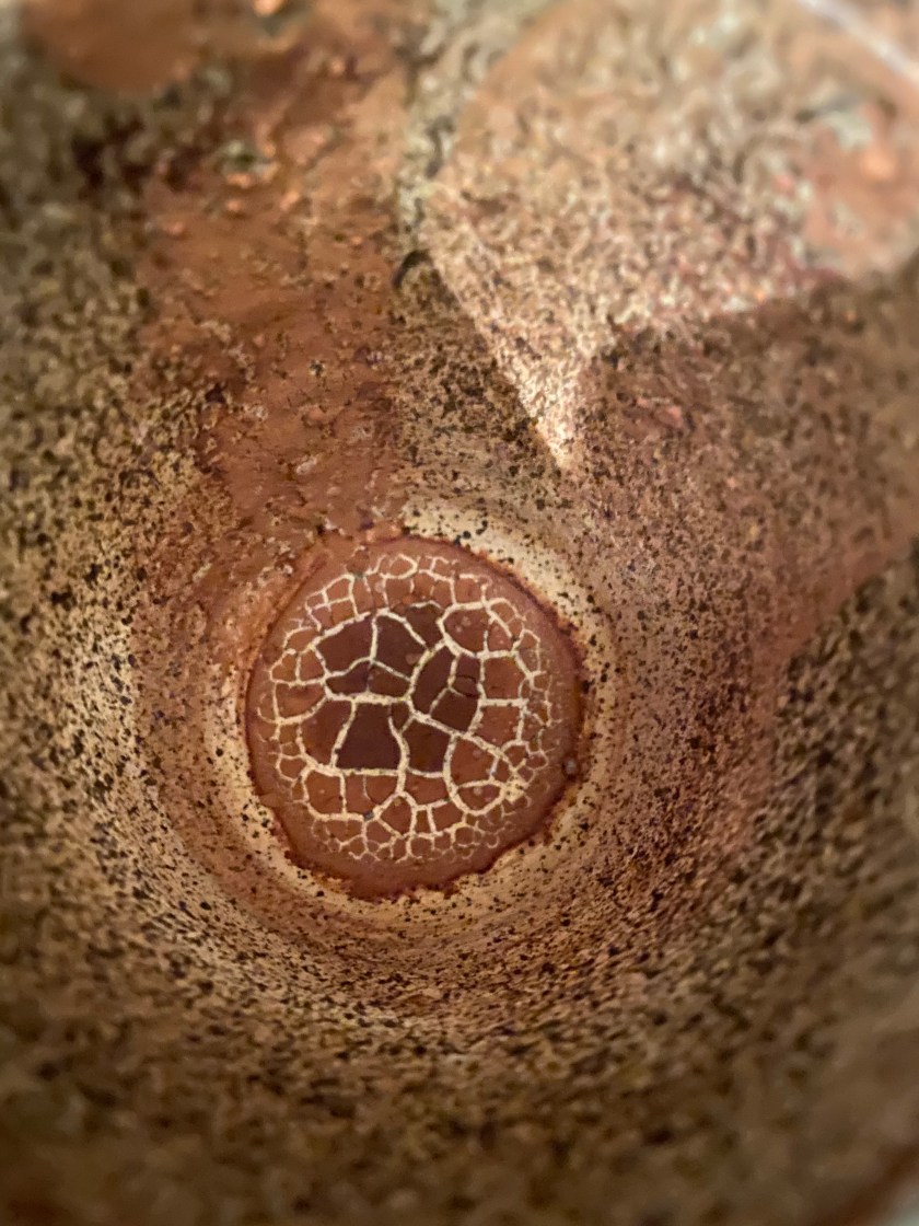
What does this have to do with the mysterious shapes at the bottom of my cacao mug? Well, in looking more closely at the full shape, I now see a spherical shape that resembles a coronavirus. Compare with this image of SARS-CoV-2:

I found the SARS-CoV-2 image in a May 4th, 2020 article from Cornell by Krishna Ramanujan: Structure of COVID-19 virus hints at key to high infection rate. The news article reviews some findings from a May 1st, 2020 paper in the Journal of Molecular Biology: Phylogenetic Analysis and Structural Modeling of SARS-CoV-2 Spike Protein Reveals an Evolutionary Distinct and Proteolytically Sensitive Activation Loop.
An interesting quote from this paper is:
Structural modeling of SARS-CoV-2 S reveals a proteolytically sensitive loop
The alignment in Figure 3(b) shows a four-amino-acid insertion 681PRRA684, as well as a conserved arginine corresponding to R685 at the S1/S2 site of the SARS-CoV-2. This insertion, which appears to be unique among lineage B betacoronaviruses, suggests a differential mechanism of activation for the SARS-CoV-2 compared to other SARS-CoV and SARS-like BatCoV.
and in the Discussion section:
In this study, we show the presence of a distinct insert that maps to the S1/S2 priming loop of the SARS-CoV-2 spike protein and is not shared with SARS-CoV or any SARS-related viruses in Betacoronavirus lineage B.
